Essential Takeaways
 Breast health needs shift over time, so your care should evolve with you.
- Age-specific screening helps catch concerns earlier and more accurately.
- Hormonal changes influence breast tissue and risk levels throughout life.
- Lifestyle choices play a meaningful role in long-term breast wellness.
- Personalized care plans support both prevention and peace of mind.
Why Personalized Breast Care Matters
Your body is not static, and your breast health shouldn’t be either. From adolescence through menopause and beyond, your breasts respond to hormonal changes, lifestyle factors, and aging in ways that require attention and flexibility. A one-size-fits-all approach can miss important signals. That is why personalized breast care is so important.
When you understand breast health by age, you are better equipped to notice changes, ask the right questions, and follow breast screening recommendations that fit your life stage. Personalized breast care approaches are not just about reacting to problems, but should also focus on building preventive breast health strategies that evolve with you.
Building Awareness in Adolescence & Early Adulthood
In your teens and early twenties, your breasts are still developing. Hormonal fluctuations can lead to tenderness, lumpiness, or asymmetry. Most changes during this stage are benign, but learning what is normal for your body is essential. Age-appropriate self-exams can help you become familiar with your breast tissue and develop a baseline with which you can compare in the future.
Routine clinical breast exams may be offered every one to three years for women in their twenties. This supports early detection breast cancer efforts without unnecessary testing.
Lifestyle and breast wellness habits also begin here. Maintaining a healthy weight, exercising regularly, and limiting alcohol use can influence your long-term risk. Research from the National Breast Cancer Foundation shows that about 13% of women in the U.S. will develop breast cancer in their lifetime, making early awareness especially valuable.
Staying Proactive in Your Premenopausal Years
As you move into your thirties and forties, your breast tissue may become denser. This can make lumps harder to detect and may affect imaging results. Hormonal cycles can still cause changes, but the focus now shifts toward more structured preventive breast health strategies.
Breast screening recommendations become more defined during this stage. Many providers suggest starting annual or biennial mammograms around age 40, depending on your risk factors. The U.S. Preventive Services Task Force recommends biennial screening mammography for women aged 40-74 to reduce mortality risk.
If you have a family history of breast cancer or genetic risk factors, your provider may suggest earlier or additional imaging, such as an MRI. Personalized breast care approaches allow your plan to reflect your personal risk, not just your age.
This stage of women’s health by age also calls for consistency. Monthly self-checks and regular clinical visits help you stay aware of subtle changes. Breast care tips, such as tracking your cycle, can help you distinguish between hormonal shifts and conditions that need evaluation.
Adapting to Change in Your Postmenopausal Years
After menopause, your hormone levels stabilize, and breast tissue often becomes less dense. While some risks decrease, others increase with age. This makes continued screening and vigilance essential.
Breast screening recommendations typically continue with regular mammograms well into your seventies, depending on your overall health. Early detection of breast cancer efforts remain important, as most breast cancer cases occur in women over 50. Recent data indicate that the median age of breast cancer diagnosis is 62.
Physical changes are also common, such as sagging or differences in texture. While these are often normal, any new lump, abnormal skin changes, or nipple discharge should be examined by your healthcare provider.
Lifestyle and breast health remain equally important at this stage. Bone health, nutrition, and physical activity all play a role in your overall well-being and can influence cancer risk. Personalized breast care means looking at your health holistically, not in isolation.
The Role of Personalized Screening & Risk Assessment
 No matter your age, your personal and family history matter. Genetic factors, reproductive history, and lifestyle choices all shape your risk profile. Personalized breast care approaches combine these elements to create a plan that works for you.
Preventive breast health strategies may include earlier screenings, supplemental imaging, or more frequent checkups. They may also involve conversations about diet, exercise, and hormone therapy. This individualized approach improves outcomes and helps you feel more in control of your health.
Take the Next Steps in Your Breast Health Care
You deserve care that sees you as an individual, not a statistic. If you are ready to build a personalized plan that reflects your age, risk factors, and goals, our team at Raleigh Gynecology & Wellness is here to help. Schedule your appointment today and move forward with confidence in your breast health.
 Changing certain habits can have a meaningful impact on long-term breast health.
 Self-exams for breast health are one of the most important ways to become familiar with how your body normally looks and feels. Set aside time once a month to check for any changes, such as lumps, changes in skin texture, or unusual tenderness.
 You do not need to adopt all these habits at once. Begin with a daily walk, or add more vegetables to your dinner plate. In time, these small steps become part of your routine.
 Spotting and mild cramping are common in the first days and weeks after
 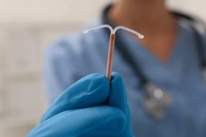 As you move through the first month, post-IUD symptoms often continue to improve. Cramping may occur occasionally rather than daily, and spotting may become lighter.
 Learning what changes and symptoms are common after IUD placement can help you approach recovery with greater peace of mind. While cramping, spotting, and cycle changes are common parts of healing after IUD insertion, you should never feel like you have to manage concerns alone.
 Your experience with
 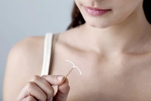 When you arrive for your IUD consultation and procedure, your provider will typically begin by reviewing your medical history and discussing your comfort preferences. You may be offered pain management options such as local anesthetic or pre-procedure medication depending on your clinic.
 A little planning ahead can make your first
 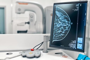 Not knowing what to expect is often a major source of
 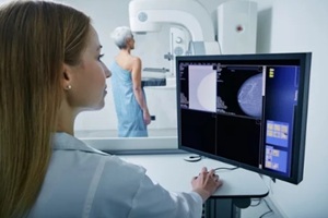 Preparing for your first mammogram is not just about checking off another medical appointment. It is about investing in your health and giving yourself the attention and care that you deserve.