 Cancer is unpredictable; some people may be more prone to it due to genetics, while others develop it in relation to lifestyle habits such as smoking. Both innate genetic expression and everyday decisions come together to impact breast health, which can be a good thing because it means that individuals can make smart choices to decrease their chances of getting cancer.
One of the best things anyone can do to reduce their cancer risk (besides getting regular mammograms!) is to incorporate regular exercise into their weekly routine. For those at risk of breast cancer (and that is both men and women!), exercise can support breast health and reduce cancer risk at the same time.
There are multiple reasons for this association between moving the body and limiting cancer risk, from how insulin is processed to the role of body fat in estrogen production. Whether a person is still in their teens or approaching retirement, it is useful to understand how exercise can support breast health and decrease cancer risk.
The Exercise-Insulin Connection
There are many reasons why exercising can reduce the risk of breast cancer. One of the most salient aspects is how exercise interacts with insulin. Insulin is produced by the pancreas in order to control how much sugar is present in the blood.
Exercise helps to regulate the insulin production cycle, reducing the development of insulin resistance. This is an essential connection, because insulin resistance is known for its correlation with breast cancer.
It can disrupt hormones, and imbalanced insulin can promote increased cell production. When more cells are being produced, there is a greater likelihood that one or more of them will replicate incorrectly, leading to cancerous growth. By exercising, individuals can keep their insulin levels stable to reduce this risk.
Moderating Estrogen with Activity
Another reason that exercise is correlated with a lower breast cancer risk is because regular movement helps to regulate estrogen production. Estrogen is associated with the tissue growth within the breast, so excess estrogen can lead to rapidly duplicating cells in the breast tissue.
As with insulin, cells that replicate quickly mathematically increase the likelihood of a cell developing incorrectly. Exercise decreases the amount of sex hormones circulating in the body, altering the ratios of specific estrogens (those that contribute to breast cancer development and those that protect against it).
Lowering Body Fat
Most people know that exercise can help them burn calories and, by extension, fat. Lowering body fat is a great way to decrease cancer risk, as fat is a primary producer of estrogen.
While exercise can improve estrogen levels, as noted previously, this impact is compounded by simultaneously decreasing fatty tissue in the body. As an added benefit, lowering the ratio of fat (and especially visceral fat around the organs) has other health benefits beyond cancer risk reduction!
Boosting Immune Function
The many cells within the immune system are the body’s innate defenders, and they can target cancerous cells in the earliest stages, as long as they can find them. Exercise facilitates this process by moving lymph throughout the body.
Lymph is the fluid within the body that carries important immune protectors, such as white blood cells, throughout the body’s tissues. Because of the forces exerted during exercise that get the heart pumping and the muscles contracting, lymph is pushed throughout the body at a higher rate than when sedentary.
This helps more immune cells circulate to even the far reaches of the tissues, where they can identify (and destroy) cancerous cells more efficiently.
Attitude and Inspiration
While exercise has plenty of tangible physical benefits, it also offers some less quantifiable advantages for patients seeking to avoid breast cancer development. When people exercise, their mood tends to improve, and over time, developing one health habit in the form of exercise can contribute to other smart choices.
Taking care of oneself is a series of decisions over an entire lifetime. Therefore, those who exercise are more likely to make smart health choices in other areas, such as regularly scheduling mammograms and actively participating in their healthcare. This helps to catch cancer early while it is still highly treatable.
Embrace Exercise as Part of Breast Cancer Prevention
 Some components of breast cancer development are found in a person’s genes, which is why regular testing is so important. However, other contributing factors are left in the hands of individuals, which is an empowering thought!
By choosing to incorporate exercise into your weekly routine, you can target multiple elements correlated with an increased breast cancer risk to mitigate your odds.
One smart choice among these decisions is to schedule your regular mammograms so any issues can be caught early. Contact Raleigh Gynecology and Wellness today to schedule your mammography scan!
 Oxytocin is most widely known as the “happy” hormone, and it impacts not just mood but also pregnancy-related behaviors such as milk production.
 Studies are currently reporting that even their
 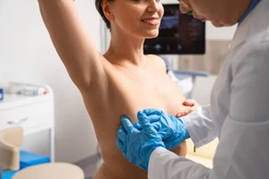 Regular doctor visits are essential for maintaining well-being and good health. However, did you know that some examinations are possible at home? Women should regularly conduct
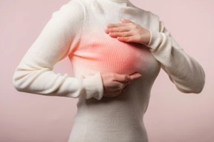
  Be sure to feel the areas around the collarbone, ribs, and armpit, as breast cancer does not solely develop in the meaty portion of the breast near the nipple.
 Breast self-examinations are the first line of defense for women to catch potential breast cancer early. The good news is that many lumps and bumps are normal, or benign, meaning they will not become cancer.
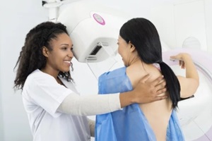
 In truth, more than 80% of suspicious lumps are first discovered on mammograms rather than noticed by women doing self-checks.
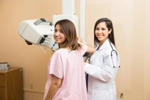 No breast cancer screening methods lower the actual risk of tumors developing in the first place. Mammograms excel at pinpointing cancers long before physical symptoms arise. This allows treatment while cells are still localized and non-aggressive.
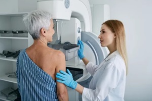 A
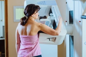
A 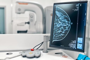 A normal (or negative) mammogram result means the radiologist did not identify any cancer or other breast abnormalities. Benign findings such as cysts or calcifications may appear, but they are not dangerous.
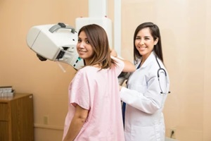 Biopsy