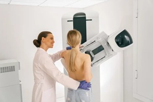
 It can be nerve-wracking to discover that the results of your latest mammogram were abnormal, prompting a visit for additional testing. However, an abnormality found in your results doesn’t necessarily mean that you have breast cancer. While mammography technology has come a long way over the last decade, it is not impervious to mistakes.
In the U.S., about 10 percent of mammograms result in call-backs for further testing. However, of these mammograms, just 7 percent lead to a cancer diagnosis. If your recent mammogram detected an abnormality, here’s what you need to know.
Understand Your Mammogram Report
Knowing how to read your mammogram report is important for better understanding what your results actually mean. When the results of a mammogram are analyzed and reported, the radiologist will categorize the results based on a standard numbered system. The system, known as the Breast Imaging Reporting and Data System (BI-RADS), categorizes mammography results based on numbers 0 to 6.
Category 0 – Incomplete
A result of category 0 means that the radiologist may have detected what they expect to be an abnormality, but the results were not clear, and additional imaging is needed for a more accurate diagnosis.
Category 1 – Negative
This is considered a normal test result, meaning the breasts appear symmetrical with no distorted structures, masses, or suspicious calcifications.
Category 2 – Benign Finding
A benign, non-cancerous, finding is considered a negative test result. The radiologist may have found a benign calcification, lymph node, or mass in the breast that is not cancer.
Category 3 – Likely Benign Finding
Findings in this category indicate a benign discovery, typically with a less than 2 percent chance of it being cancer.
Category 4 – Suspicious Abnormality
While these findings do not always result in a breast cancer diagnosis, suspicion will often dictate the need for a biopsy. Findings in this category are further broken down based on the likelihood of cancer, ranging from low likelihood to high likelihood.
Category 5 – Highly Suggestive of Malignancy
 If you get a Category 5 result, appropriate action should be immediately taken. The findings will typically look like cancer and have a high chance of cancer, usually at least 95 percent. With this result, a biopsy is strongly recommended.
Category 6 – Proven Malignancy
A Category 6 result is generally reserved for findings on imaging that have already proven to be cancer from a previous biopsy. Immediate action should be taken to determine the best course of action, and additional imaging may be used to see how the cancer responds to treatment.
What to Expect at Your Follow-Up Appointment
If you received an abnormal result on your mammogram, you’ll likely be asked to come in for a follow-up appointment. The type of additional testing recommended to you by your provider will depend on the categorization of your test results, the density of your breast tissue, and other relevant factors.
A diagnostic mammogram may be recommended to get more accurate photos of the areas of concern. This imaging is similar to a routine mammogram but focuses on the mass or other abnormality seen in the previous tests. A radiologist will then be contacted to review the new images.
If more detailed imaging is needed after receiving abnormal results on a mammogram, a breast ultrasound or breast MRI may be recommended. An ultrasound of the breast uses sound waves to generate pictures of the inside breast tissues and structures.
This non-invasive procedure can be useful for differentiating between fluid-filled masses, such as benign cysts, and solid masses, which could indicate cancer. A breast MRI may be recommended for women at high risk for breast cancer or to evaluate the extent of breast cancer after a diagnosis.
During this procedure, you’ll lie face down on a padded table with your breasts positioned in the appropriate openings. The table is then slid into an MRI machine where detailed images are taken using radio waves and magnets. A contrast agent may be injected to achieve better visualization.
In some cases, you may need a biopsy to check for cancer. This procedure involves removing a small piece of the tissue to be examined under a microscope. Several different types of biopsies exist, such as those done with a small, hollow needle and those completed through a small cut in the skin.
Call Raleigh Gynecology & Wellness for Breast Care
 Choosing the right technology and the right care provider when approaching breast cancer can make all the difference in your health outcome. At Raleigh Gynecology & Wellness, we offer the latest advancements in breast cancer screening.
Genius 3D Mammography exams can find 20 to 65 percent more invasive breast cancers compared to traditional 2D mammograms. Contact our office today to learn more about this exam or to discuss any concerns you may have about your breast health.
