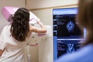
 The mention of a “mammogram” often sparks anxiety for women worried about potential breast cancer diagnosis. However, breakthroughs in breast imaging technology over the past century have transformed early detection and reduced deaths from this disease.
Learning about the ongoing progress toward catching breast tumors at more treatable stages can help you appreciate the fundamental role screening plays in safeguarding health.
Laying the Groundwork for Mammography
German surgeon Albert Salomon captured the first X-ray image of mastectomy specimens in 1913 to analyze how cancers spread to lymph nodes. This primitive use of radiology to visually assess excised breast tissue established foundations for the field of mammography.
In the 1930s, several important discoveries moved clinical imaging of actual breasts forward. Surgeons began X-raying the breasts of living patients to aid in diagnosing palpable lumps found during exams. Radiologists could then study these images to determine if tumors appeared present alongside any observed calcifications.
Standardizing Breast Imaging Techniques
Early mammogram image quality and breast tissue visibility varied widely depending on the equipment used. So, pioneers such as radiologist Robert Egan developed compression techniques to flatten breasts. This made abnormalities easier to spot while allowing lower radiation doses to capture clearer pictures.
Such incremental technical refinements eventually paved the way for dedicated mammography devices expressly designed for breast imaging. Regulations also emerged requiring minimum performance standards, rigorous quality control programs, and imaging staff training protocols.
Systematizing efforts in properly executing and accurately interpreting mammograms formed foundations for widespread screening. And the capacity to reliably detect subtle cancers before they became palpable or spread proved essential to dramatically improving breast cancer survival rates.
Harnessing the Life-Saving Potential of Mammograms
Early detection to catch non-palpable breast tumors offers women diagnosed with cancer much a better prognosis compared to more advanced stages of disease. However, a debate ensued around using mammography to screen seemingly healthy women rather than only diagnosing symptomatic patients.
Concerns over cost-effectiveness, radiation hazards, and false positive rates initially suppressed enthusiasm for screening exams. That reluctance to adopt population-level testing reversed thanks to statistically persuasive clinical studies from the 1970s onward.
Research by the Health Insurance Plan of New York involving over 60,000 patients demonstrated a 30% breast cancer mortality reduction. The Swedish Two-County Trial and other projects validated that early detection through universal mammographic screening programs could lower death rates by at least 25%.
These compelling results sparked the widespread adoption of regular breast cancer checks via low-dose X-ray mammography. Data confirms that screening remains our most powerful weapon for combating this disease.
Ongoing Refinements Expand Capabilities
 While film screen mammograms still play a valuable clinical role today, digital advancements now enable greater precision. Full-field digital mammography (FFDM) converts X-rays into electronic images using detectors rather than old-fashioned film.
This innovation opened doors for new techniques, such as tomosynthesis. Here, multiple image “slices” taken from different angles get reconstructed into highly detailed 3D visualizations. This fights a long-standing mammography challenge involving overlapping tissue obscuring potential trouble spots.
Recently approved contrast-enhanced mammography (CEM) offers another way to distinguish benign from malignant cases. Special dyes injected intravenously before a scan help suspicious areas “light up” clearly.
CEM produces fewer false positives than standard scans, reducing the need for biopsies and the associated stress for women who are worried they might have cancer.
Ultrasound and MRI Scans Add Supplemental Detection Power
Screening mammograms are the first line of defense in routine breast cancer checks. But supplemental ultrasound and MRI evaluations provide complementary strengths by revealing tumor characteristics using soundwaves or magnetic fields.
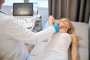
Handheld ultrasound wands pressed against breast skin supply valuable anatomical details often obscured on X-ray films. Additionally, Doppler techniques spotlight blood vessel development that could supply the fuel for possible malignant growths.
Finally, MRI platforms offer unmatched sensitivity when uncovering disease sites mammography or ultrasound might miss.
Artificial Intelligence Skyrockets Accuracy
Complex computer algorithms that imitate human evaluation of breast images are among the most significant breakthroughs on the horizon. Artificial intelligence (AI) will skyrocket diagnostic accuracy, efficiency, and accessibility into new dimensions.
Deep learning neural networks can zero in on highly subtle cues that human eyes evaluating hundreds of images per shift easily overlook due to mental fatigue. Automating these tasks frees radiologists to focus more on delivering compassionate patient care.
Furthermore, broad data sharing empowers AI tools to spot patterns by scanning millions of mammogram images rather than limited samples from any institution.
This improves diagnostic accuracy and makes this invaluable expertise available to all by connecting underserved regions to cutting-edge diagnostic infrastructure.
But effectively, personalized breast care still requires human direction. AI generates probability scores for suspicious findings, but only doctors can gauge whether the risks of a biopsy make sense over waiting to see how the situation progresses.
The Road Ahead: Precision Screening
 Despite fantastic progress in lowering breast cancer deaths through early detection, every woman’s ultimate hope is for the disease not to develop at all. So, innovations centered on more strategic screening aim to expose fewer patients to unnecessary radiation risks or false positive scares.
Precision health models strive to pinpoint individuals facing elevated risk rather than adopting blanket testing policies. Specific genetic markers, breast density patterns, or hormonal signatures may clue clinicians into higher threat categories warranting extra attention.
Raleigh Gynecology & Wellness: Your Ally in Early Detection
Raleigh Gynecology & Wellness understands the essential role thoughtful, compassionate screening plays in giving patients the best chances against breast cancer. We continuously monitor which exam strategies and frequencies make the most sense for your risk profile and personal health goals.
So turn to Raleigh Gynecology & Wellness for the perfect partnership blending cutting-edge cancer detection with unwavering emotional support. Contact us at (919) 636-6670 or online to discuss what breast wellness strategies are right for you.
